Sunday, December 14, 2014

Cranial Technique and craniosacral therapy and Applied Kinesiology


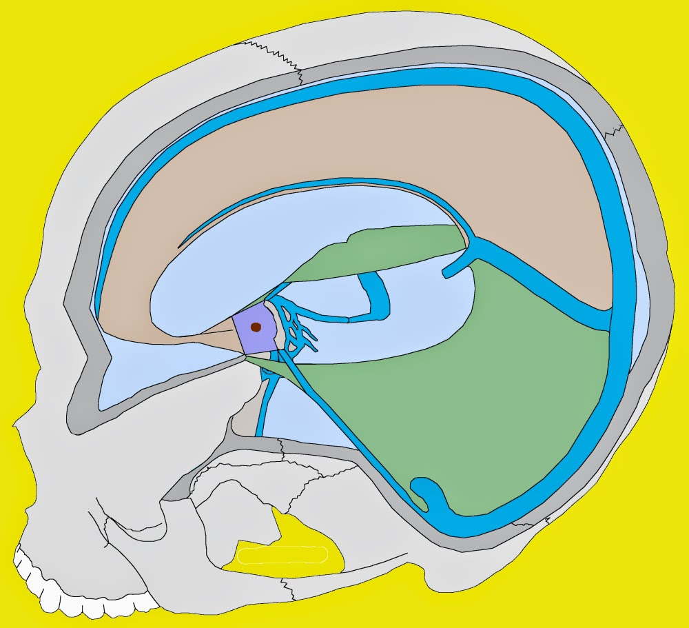
For several decades Dr. George J. Goodheart, Jr. produced Monthly AK Research Tapes -- LOADED with exceptional insights for the holistic physician. Here are the "Highlights" of Tape 4, Side 1 -- (with a few "illustrative pictures")

"Challenge of Cranial Faults
...
A. Sign of internal or external rotated frontal is weakness of anterior neck flexors. 


 B. Internal frontal bone signs.

  1. Wide nares on one side.

  2. Narrow orbit on other side.

  3. Super orbital notch soreness.

 C. External frontal signs.

  1. Wide orbit

  2. Painful eyeball on wide orbit side.

  3. Painful cheek bone on opposite side.

 D. These external signs may be present without anterior neck flexor weakness.

 E. Procedure for challenging internal frontal. Example: Wide nares, narrow orbit side.


  1. Patient is in supine position.

  2. Put pressure on malar bone.

  3. Press medially toward base of nose with 4 or 5 lbs. pressure.

  4. If there is a partial internal frontal bone rotation, the anterior neck flexors will show strength, but when pressure is applied to the malar bone as described above, this will cause an immediate weakness of the anterior neck flexors.

 F. Correct internal frontal bone in usual way.

  1. Go to side of wide nares, narrow orbit using roll out type pressure on the alveolar process.

  2. Go past teeth into pterygoid pocket and press footward.

  3. Get on pterygoid process on the opposite side of press upward.

 G. Re-challenge malar arch and anterior neck flexors will not blow.

 H. External frontal bone rotation.

  1. Wide orbit with no change in nares.

  2. The wide orbit side has painful eyeball.

  3. Narrow orbit side has painful cheek bone.

 I. External frontal bone challenge.


  1. Go to narrow orbit side.

  2. Grasp upper molar teeth and pulls downward with 4 or 5 lbs. pressure, (if the patient has dentures, try to grasp gums in that area).

  3. Retest anterior neck flexors.

  4. If partial external frontal bone rotation, flexors will go weak.

  5. Correct external frontal bone as follows:

  a. Press lateral to cruciate ligament on hard palate on narrow orbit side, press toward vertex of skull.
b. Check eyeball pain, pressing on cruciate ligament in direction that eliminates it.


 J. Use in difficult cervical and whiplash injuries."


Applied Kinesiology and Traditional Chinese Medicine or Acupuncture


More Clinical Wisdom (about Acupuncture and AK) offered by Dr. Goodheart in his Monthly AK Research Tapes.

(Doctors around the world remember Dr. Goodheart spe...aking with this kind of detailed clarity about all these issues when they spoke with him...inexhaustible GENIUS!)

"Temperature difference in Acupuncture.
A. Inner aspect of knee is related to liver. 

 B. Outer aspect of knee is related to stomach.

 C. Patella aspect of knee is related to spleen.

 D. Lateral portion of lower limb, starting at head of fibular to lateral maleolus is related to gallbladder.

 E. Medial aspect of leg from the Achilles tendon to the os calcis is related to the kidney.

 F. Midway on the radial ulnar aspect of arm on the palmar surface is related to the paracardium or circulation sex, (the P-4 or H-4 area).

 G. On the volar aspect of the arm from the olecranon process to the mid-portion of the transverse ligament, is related to the thyroid gland.

 H. On the base of the metacarpal to the tip of the little finger on the little finger side is the area associated with the small intestine.

 I. At the point L-1 to L-11, or what the Chinese call the "Three mile point." On the radial side of the arm at the elbow, the size of one half dollar, is the area associated with the large intestine.

 J. The mid-aspect of the lower limb at point K-7 is the area for the kidney or psoas.

 K. These areas will be hot to the touch if the meridian is full.

 L. These same areas will be cool if the meridian is empty or deficient.

A quick review of the above.
 
A. Bladder or sacrospinalis, B-59 to B-60, (medial aspect of Achilles tendon).

 B. Liver or pectoralis major sternal division, L-8, (medial aspect of knee).

 C. Stomach or pectoralis major clavicular division, S-34 to S-36, (lateral aspect of knee).

 D. Gall bladder or popliteus muscle, G-33 to G-39, (lateral aspect of lower leg).

 E. Spleen lower and middle trapezius, pancreas, latissimus dorsi, St-9, (adjacent to the patella).

 F. Heart or subscapularis, H-7, (on the palmar surface of the hand, little finger side).

 G. Note: Correlate the above areas with Chinese pulse evaluation, alarm points, and muscle testing."

(MORE)

"Acupuncture

A. Running a meridian from beginning to end turns muscle on.

 B. Running a meridian from the end to the beginning, turns a muscle off.


Example:

1. Running kidney meridian from K-1 to K-27, will turn on psoas muscle. 

 2. Running a kidney meridian from K-27 to K-1 will turn off psoas muscle with the exception:

 a. When too much energy exists in a meridian, that meridian will not turn off. (Plus meridian).

 C. Conception vessel works in reverse.

 1. When running CV-24 to CV-1, a positive muscle response is elicited.

 a. Any muscle will turn on that is associated with a cranial fault.

 2. Conception vessel locks cranial fault in corrected position. Also locks upper cervical and occipital faults, (atlas, axis, and 3rd cervical).

 D. Governing vessel.

 1. Starts at coccyx tip, runs over the head on midline, over the nose to upper lip.

 a. Follows usual course, running forward turns muscle on, turning backward turns muscle off.

 2. Governing vessel is associated with sacral faults.

 3. Does not lock pelvis as unit, (posterior ilium, posterior ischium, etc.).

 4. It will only lock a sacral fault, respiratory spinal fluid in nature.

 5. A sacral respiration fault cannot be reversed, once the governing vessel has been locked.

 E. A cranial respiratory fault cannot be reversed once the conception vessel has been locked."


The latest information and research evidence supporting the combination of manual muscle testing for the analysis of Meridian System Disorders is here:
Chapter 10 -- Applied Kinesiology Essentials: The Missing Link In Health Care

https://www.thegangasaspress.com/


Saturday, December 6, 2014

Craniosacral treatment with Applied Kinesiology

For several decades Dr. Goodheart produced Monthly AK Research Tapes -- LOADED with exceptional insights for the holistic physician. Here are the "Highlights" of Tape 5, Side 1--
(courtesy Dr. Paul White, DC, DIBAK):


Challenge of Cranial Faults
A.   Sign of internal or external rotated frontal is weakness of   anterior neck flexors.
B.   Internal frontal bone signs.
     1. Wide nares on one side.
     2. Narrow orbit on other side.
     3. Super orbital notch soreness.
 
C.   External frontal signs.
     1. Wide orbit
     2. Painful eyeball on wide orbit side.
     3. Painful cheek bone on opposite side.
D.   These external signs may be present without anterior neck      flexor weakness.
E.   Procedure for challenging internal frontal. Example: Wide      nares, narrow orbit side.
     1. Patient is in supine position.
     2. Put pressure on malar bone.
     3. Press medially toward base of nose with 4 or 5 lbs.      pressure.
     4. If there is a partial internal frontal bone rotation,      the anterior neck flexors will show strength, but when pressure is applied to the malar bone as described above, this will cause an immediate weakness of the anterior neck flexors.
F.   Correct internal frontal bone in usual way. 


     1. Go to side of wide nares, narrow orbit using roll out      type pressure on the alveolar process.
     2. Go past teeth into pterygoid pocket and press footward.
     3. Get on pterygoid process on the opposite side of press      upward.
G.   Re-challenge malar arch and anterior neck flexors will not blow. 


H.   External frontal bone rotation.
     1. Wide orbit with no change in nares.
     2. The wide orbit side has painful eyeball.
     3. Narrow orbit side has painful cheek bone.
I.   External frontal bone challenge.
     1. Go to narrow orbit side.
     2. Grasp upper molar teeth and pulls downward with 4 or 5              lbs. pressure, (if the patient has dentures, try to           grasp gums in that area).
     3. Retest anterior neck flexors.
     4. If partial external frontal bone rotation, flexors will              go weak.
     5. Correct external frontal bone as follows:
          a. Press lateral to cruciate ligament on hard palate                   on narrow orbit side, press toward vertex of skull.
          b. Check eyeball pain, pressing on cruciate ligament                   in direction that eliminates it.
J.   Use in difficult cervical and whiplash injuries."

 
Find out more about the Applied Kinesiology
Approach to Cranial Disorders at: